One of science’s greatest leaps was the discovery of X-rays. It took the scientific world, and the public, by storm when it was first introduced in 1895 — and with good reason. The development of the X-ray led to numerous breakthroughs in a variety of industries, not just medical.
How did it all start? Well, you’re in the right place to find out.
Who Discovered X-ray
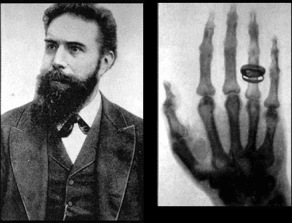
X-rays were discovered by Wilhelm Conrad Roentgen, a German physicist. He made this groundbreaking discovery on November 8, 1895, while conducting experiments with cathode-ray tubes in his laboratory at the University of Würzburg.

Accidental Beginnings
It’s the year 1895, and a German physicist named Wilhelm Conrad Roentgen is working in his laboratory. He’s experimenting with cathode-ray tubes, which produce a kind of invisible light. One day, while he’s working, he notices something unexpected.
There’s a special kind of screen in the room that glows when hit by these invisible rays. Roentgen realizes that something unusual is happening. To investigate, he covers the tube with heavy black paper, and to his surprise, the screen still glows. This means the invisible rays can pass through solid objects!
This accidental discovery piques Roentgen’s curiosity, and he decides to explore further. He places different objects in the path of these invisible rays and notices that they create shadows of the objects on the screen. This is like holding your hand in front of a flashlight, and you see a shadow on the wall.
To make it even more intriguing, Roentgen asks his wife to place her hand in the path of these invisible rays. What appears on the screen is a detailed shadow of the bones in her hand, complete with her wedding ring. This is the birth of X-ray imaging!
X-rays have come a long way since then and are now used by companies for multiple procedures using technology like that offered by Maven Imaging — but this original discovery is what started it all!
Evolution of X-ray Technology: A Timeline of Milestones and Advancements
Here’s a brief timeline highlighting key events in the growth and history of X-rays:
- 1895: Wilhelm Conrad Roentgen accidentally discovers X-rays while experimenting with cathode-ray tubes at the University of Würzburg.
- 1896: Roentgen publishes his discovery, and X-rays are quickly adopted for medical use. The first medical X-ray is taken, showcasing the bones of a human hand.
- 1896: X-rays are used in medical settings for the first time by surgeons to locate bullets in wounded soldiers during the Balkan War.
- 1896-1897: Thomas Edison investigates X-rays and develops a fluoroscope, an early X-ray imaging device.
- 1901: Roentgen is awarded the Nobel Prize in Physics for the discovery of X-rays.
- 1913: X-ray crystallography is developed by Sir William Henry Bragg and his son William Lawrence Bragg, allowing the study of crystal structures.
- 1922: The first commercial X-ray machine is introduced by General Electric.
- 1930s: Introduction of the X-ray tube that allows adjustable X-ray dosage.
- 1940s: Widespread use of X-rays in medical diagnostics becomes a standard practice.
- 1950s-1960s: Advancements in X-ray technology, including the development of computed tomography (CT) scanning.
- 1970s: Introduction of digital imaging, moving from traditional X-ray film to electronic detectors.
- 1980s: Magnetic Resonance Imaging (MRI) technology becomes a powerful tool in medical imaging alongside X-rays.
- 1990s: Introduction of digital radiography, further improving image quality and accessibility.
- 2000s: Continued advancements in X-ray technology, with the rise of digital radiography and improvements in image processing and computer-aided diagnosis.
- Present: X-rays remain a fundamental tool in medical diagnostics, as well as in various industries such as manufacturing and security. Ongoing research and technological developments continue to enhance the capabilities and applications of X-ray imaging.
This timeline provides a snapshot of the major milestones in the growth and evolution of X-ray technology. The field continues to progress, contributing significantly to both medical and industrial applications.
Further Discoveries
Through further experimentation, Roentgen discovered that these rays could pass through human tissue but not through bone or metal objects. This led to one of the first iconic images produced by X-ray, as Roentgen somehow convinced his wife, Bertha, to present her hand before the rays. What emerged was an image of her skeletal hand with her wedding ring attached.
This led to many swift and exciting developments in the scientific community, namely due to its accessibility. Due to its relationship to cathode tubes (which were well understood at the time), many scientists could begin conducting their own research into X-rays. This excitement spread from the scientific community through to the public.
An Exciting New Development
Newspapers and magazines communicated news of this invention to the broader public, and understandably, intrigue abounded. The notion of an invisible ray that could pass through solid matter seemed like something from a fictional story rather than scientific fact. There’s no doubt that some of these reports exaggerated the real potential of the X-ray, but the excitement from the public was clear.
This enthusiasm was, in most cases, justified. Within just a month, surgeons throughout Europe and the United States began to use medical radiographs to assist them when treating patients.
In June of 1896, just six months after Roentgen first announced his incredible discovery, X-rays were used by military medics to locate bullets in wounded soldiers. In no time, X-rays had become a staple of medical treatment.

Modern Applications: Where We Are Now
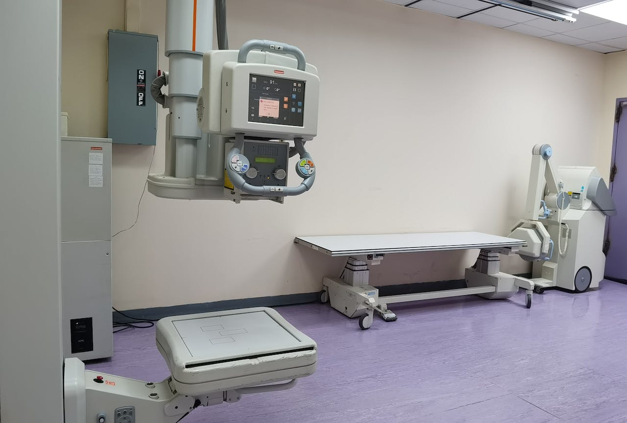
Although over a hundred years have passed since Roentgen discovered X-rays, radiography hasn’t changed enormously. The original principles still apply, capturing a shadowed image to improve visibility. The main difference now is the sophistication, clarity, and sensitivity of the images produced.
The incorporation of electronics and computers allowed for the replacement of film, meaning greater efficiency and automation could take place. Now, X-rays are a staple of multiple industries, including medicine and manufacturing.
From canned food to airbags, radiography has its place. We can’t know for sure where X-ray technology will go from here, but we know that it’s cemented its place in history.
Related Posts


It’s great that you talked about how one of science’s greatest leaps was the discovery of X-rays. I was watching an educational TV show yesterday and it discussed how X-ray was developed and used. According to what I’ve watched, it seems there are various services centered around X-rays now too, like nursing home X-ray services for example.