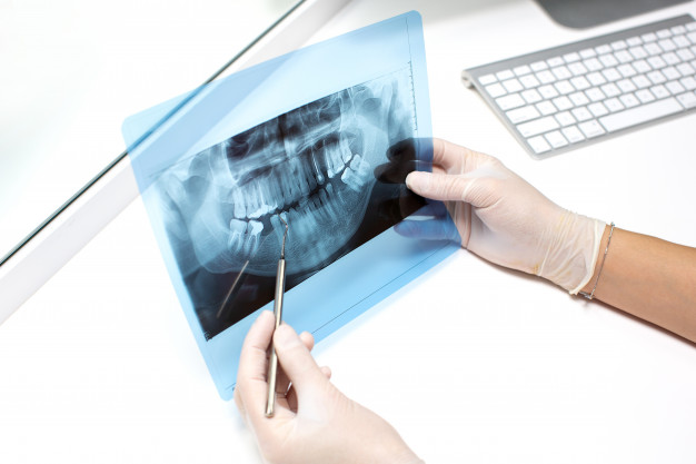
Dental radiographs are commonly known as dental X-Rays and they are often required for a dentist to make an accurate diagnosis, as he or she needs to see the jawbone structure. A radiographic image is taken with a controlled burst of X-Ray radiation, which is not harmful. The image takes into account the various densities in the patient’s mouth to give an image that a specialist can read.
Intra-Oral X-Rays
As the name suggests, the film or sensor is placed inside the mouth. Here are some of the conditions that intra-oral imaging is used:
- Detecting gum infection
- Evaluating periodontal issues
- Assessing trauma fractures to teeth or bone
- Pre-extraction evaluation
- Detecting the presence or position of unerupted teeth
- Root canal treatment
This would also be used to check the condition of recently fitted dental implants and even to assess where the best location for the titanium pins would be. If you are in need of dental treatment, the best BUPA dentist on the Gold Coast is only a phone call away, if you live in the area. Check for a reputable dentist in your location through a Google search if you haven’t got your own dentist yet. You should have a check-up every 5-6 months.
Types of Intra-Oral X-Rays
There are several types of intra-oral X-Rays:
- Bite Wing X-Rays – These are primarily used to spot decay between teeth as well as changes in bone density brought about by gum disease. This type of imaging is also used to determine the best crown location.
- Periapical X-Rays – This shows the complete tooth from the crown to the very end of the roots and are used to evaluate the root structure.
- Occlusal X-Rays – Larger images that cover the entire upper or lower section of the teeth.
When you visit your dentist and he or she feels that X-Rays are called for, the process doesn’t take long and once the images are ready, the dentist often shows the patient while explaining what this means. Click here for further reading on dental X-Rays.
Extra-Oral X-Rays
While intra-oral X-Rays actually take an image inside the mouth, the extra-oral method takes the image from outside, with the film or sensor placed on the outside of the cheek in the right position to capture the desired image. Here are a few reasons why extra-oral images are required:
- Diagnosis of skeletal abnormalities
- Treatment planning
- To monitor the progress of treatment
- Evaluation of orthodontic treatment
- Assessment of misaligned teeth
- Evaluate root length

X-Rays are a critical component for dentistry and using computer technology, clear images can be achieved, which help the dentist to evaluate, measure and monitor dental issues and treatment. Most people have had dental X-Rays at some time in their life and with a radiologist in attendance, there’s nothing to worry about with this painless experience.
While on the subject of dental treatment, we thought we should remind you of how important it is to have best oral hygiene practices, which include regular brushing and flossing after every lean and before and after sleep. You should also use an antiseptic mouthwash regularly, after eating snacks or drinking alcohol, which really helps to maintain healthy teeth and gums.
Related Posts


One thought on “Intra-Oral Radiography: All You Need To Know”
Comments are closed.